

Tissue AI
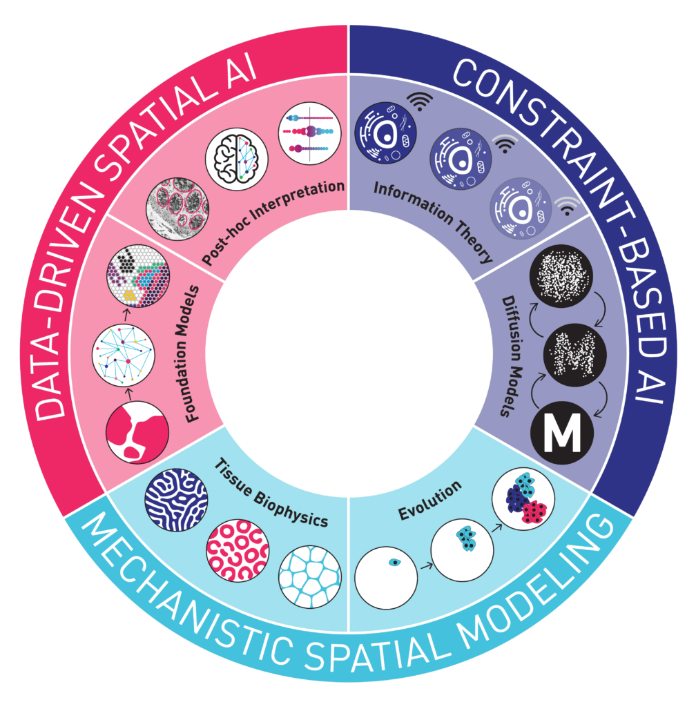
A major theme of the lab is Tissue AI, in which we use artificial intelligence and other computational approaches to understand the spatial processes within biological tissues. Our approaches combine Data-Driven Spatial AI, Constraint-Based AI, and Mechanistic Spatial Modeling. We focus on analyzing cancer image data to understand treatment response, such as how it is mediated by tumor subtype status, intratumoral heterogeneity, immune cells, and other microenvironmental features (Rubinstein, Domanskyi et al 2025, Noorbakhsh et al 2020, Mukashyaka et al 2024, Farahmand et al 2022). The lab investigates modern imaging datatypes including spatial transcriptomics (Visium, Xenium, VisiumHD, merFISH), spatial proteomics (CODEX, imaging mass cytometry), spatial long-read sequencing, and H&E/IHC . Our research team is made up of computational biologists, cancer biologists, immunologists, cancer surgeons, physicists, and pathologists, providing expertise to interrogate these problems in interdisciplinary ways. We collaborate closely with experimental biologists and oncologists to clarify and validate findings.
Our lab also uses Tissue AI to study the variations in function across normal tissues, both in human and mouse. We co-lead the Data Analysis Core for the KAPP-Sen U54 in the NIH Cellular Senescence Network. In this project we analyze H&E, spatial transcriptomics, and spatial proteomics datasets for kidney, adipose, pancreas, and placental tissues (NIH SenNet Consortium, 2023).
Cancer Ecosystems
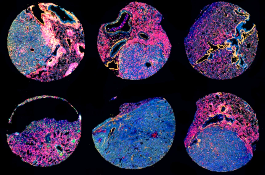
We study how intratumoral ecosystems and cellular interactions impact tumor growth and treatment response. As an example, aging is the biggest risk factor for cancer, yet it is not fully understood why. We are studying how aging changes the prevalence and interactions of cancer and immune cells, particularly in the breast and lung. In collaboration with the JAX Aging Center we are studying tumors from aged mice, allowing us to uncover spatial patterns of cells that arise in cancer during aging. Linking these patterns to patient outcomes could reveal why cancer becomes more dangerous with age and point to new ways to improve treatment for older patients (Angarola et al 2024).
We also study Cancer Ecosystems using mechanistic computational modeling approaches from a variety of imaging technologies. For example, we recently developed a method to identify cell interactions based on how proteins accumulate at points of contact between cells (Wang et al 2024). Using this approach, we discovered that interactions between two types of immune cells—T cells and B cells—within tumors are linked to better patient survival. This finding opens a promising new strategy for identifying patients who may respond well to treatment and for improving outcomes by targeting these cell interactions. We have developed and applied such approaches based on both 2D and 3D characterizations of Cancer Ecosystems (Mukashyaka, Kumar, et al 2023).
Patient-Derived Xenografts
The Chuang lab is a leader in the field of patient-derived xenografts (PDXs), a model system in which human tumors are engrafted and studied in NSG mice. Xenografts play a critical role in cancer research, as they are used in therapeutic testing to verify drug activity before embarking on a clinical trial. Since 2017, Dr. Chuang has led the Data Coordination Center for the PDX Network, a multi-institute consortium supported by the US National Cancer Institute. We are partnering with institutes including the Huntsman Cancer Institute, Baylor College of Medicine, MD Anderson, Washington University, the Wistar Institute, the University of Pennsylvania, Dana Farber Cancer Institute, Virginia Commonwealth University, the University of California-Davis, and Frederick National Laboratory, to study cancer using xenografts. Our work has produced pioneering studies on the genetic (Woo, Giordano et al 2021) and therapeutic (Evard et al 2020) robustness of xenografts for preclinical testing. Our lab also contributes to the PIVOT Consortium, an NCI project to test the efficacy of drugs for pediatric cancers using xenografts. Within these xenograft projects, our lab is especially focused on the dynamic response of cancers to treatment (Rubinstein, Domanskyii et al, 2025), as xenografts make it possible to reproducibly interrogate this process at critical moments including pre-treatment, early treatment, minimal residual disease, and the onset of resistance.
